
Nanoparticles made from bacteriophage proteins could be used in the future to treat cancer.
When developing new synthetic materials, scientists are often inspired by the variety of structures and shapes created by nature. The smallest biological objects - viruses, such as bacteria-infecting viruses, or bacteriophages - are also being targeted. Often resembling miniature alien spaceships or tiny spiders, they are everywhere around us. They are the most abundant form of life in the biosphere. With millions of them in a drop of water or a pinch of soil, it's no wonder that scientists see their benefits too. The most common application of bacteriophages is for the detection or killing of pathogenic bacteria. They are also used to develop new materials, such as human-enhanced viruses or virus components in nanotechnology. Smaller than bacteria but larger than proteins, nanoparticles are miniature natural or synthetic cages designed to carry genes, drugs or other therapeutic or diagnostic compounds. Over the past decades, scientists have created nanoparticles from a wide variety of materials such as lipids, proteins, carbon, various metals and viruses or their components. By attaching molecules to the surfaces of such particles that recognise, for example, cancer cells, damaged blood vessels or the focus of an infection, scientists hope they will deliver drugs to just the right target. Such targeting could reduce the strong side effects of drugs.
In their research, scientists at the VU Life Sciences Centre aim to create nanoparticles from a bacteriophage protein that can self-assemble into the tubular structures. By genetically fusing fluorescent molecules to this protein, they have succeeded in producing luminous nanotubes. This allowed the researchers to observe by fluorescence microscopy the delivery of the nanotubes to different types of cells: immune cells, i.e. brain macrophages (microglia) and peritoneal macrophages, and colorectal cancer cells. The nanoparticles have been shown to accumulate in the cells and are not toxic to them. We found that the microglial cells of old mice are more efficiently infiltrated by nanoparticles than those of young mice. The opposite trend was observed in peritoneal macrophages, where nanoparticles were more efficiently delivered to the cells of young mice. This suggests that ageing affects different macrophages differently in the body, leading to variable entry and removal of nanoparticles depending on age. Nanoparticles were also efficiently incorporated into colon cancer cells and accumulated in their lysosomes. By attaching an enzyme to such nanoparticles, which produces a cell-killing drug from a compound that is not harmful to the body, we plan to target the nanoparticles to cancer cells and further investigate the so-called prodrug-enzyme system we have developed.
The research by the VU LSC scientists has only just begun, but the fact that the bacteriophage protein nanoparticles enter cells and their accumulation is linked to the age of the organism from which the cells are derived is promising for their medical applications as carriers for the treatment of some diseases. Based on the results obtained in this project, we recommend the use of nanocarriers based on bacteriophage tail sheath proteins for the delivery of therapeutic molecules to cancer cells. The response of peritoneal macrophages to nanotubes suggests that age-related changes in cells lead to a poorer uptake of nanotubes based on bacteriophage tail proteins. The efficient internalisation of these nanotubes by cancer cells would lead to the development of efficient nanocarriers for therapeutic agents based on these proteins.
The research was carried out as part of the National Science Programme (NSP) "Healthy Ageing" project "Self-assembling phage proteins for targeted nanomedicine", funded by the Lithuanian Research Council. The project was carried out by Vilnius University, Life Sciences Centre researchers Vida Časaitė, PhD, Aušra Sasnauskienė, PhD, Doc. Dovydas Gabrielaitis, PhD. Vilmantė Žitkutė, PhD. Lina Saveikytė, Stud. Greta Labutytė.
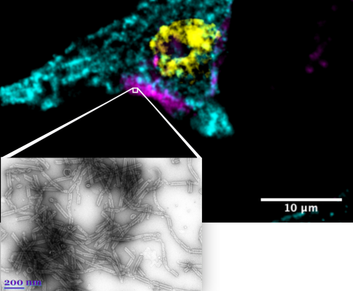
The image shows nanotubes observed by fluorescence microscopy inside a cell (top photo), where purple is the nanotubes, green is the cytoplasm of the cell, and yellow is the cell nucleus. Bottom photo shows the nanotubes as seen under an electron microscope.